第一作者:單旭晨、王帆博士
通訊作者:金大勇教授、Peter Reece博士、王帆博士
通訊單位:悉尼科技大學、新南威爾士大學
研究背景
光鑷技術已經被廣泛的應用于材料的組裝、表征,細胞內抓取和力的測量。1997年諾貝爾物理獎就是表彰Steven Chu, Claude Cohen-Tannoudji 和 William D. Phillips用單原子光鑷方法來探索原子在光學場中的共振從而對單原子限域和冷卻。2018年的諾貝爾物理獎的一半獎金用于表彰Arthur Ashkin光鑷來探索生物系統中的應用。
通常光鑷的捕獲力都是由捕獲顆粒和周圍介質的折射率差決定的,顆粒的折射率比周圍介質越大其受到的光力就越大。此外顆粒的尺寸越小,其收到的光力效果就越小。這兩個原因導致了低折射率顆粒(例如納米顆粒,納米藥物和細胞器)的光學捕獲尤為挑戰。
選擇高介電常數材料例如金屬顆粒和半導體顆粒作為光鑷的操縱探針是現階段主要的方法。但是金屬顆粒所產生的光熱效果不僅會影響光鑷操縱效率還會影響生物樣品。現階段半徑50納米金顆粒能提供的光力彈性系數是0.012 pNμm-1mW–1, 而半徑52納米的硅顆粒能提供的光力彈性系數也只有0.022 pNμm-1mW–1.
成果簡介
最近,澳大利亞悉尼科技大學(UTS)的研究者通過對材料科學,化學合成和光子物理結合,運用光學物理機制實現了對低折射率 (n=1.46) 納米顆粒的光力增強,提高為普通金納米顆粒的三十倍,并且首次實現了該納米顆粒在無折射率差環境中的光學捕獲。該項多學科交叉的研究成果相關工作以“Optical tweezers beyond refractive index mismatch using highly doped nanoparticles”為題,發表于最新一期的《Nature Nanotechnology》 上。
研究出發點
稀土摻雜的上轉換顆粒是一種可以將入射的近紅外光轉化為可見和近紅外光的新型材料。顆粒中摻雜的給體稀土離子可以吸收長波長的紅外光,將其傳遞給受體發光離子,從而發出短波長的可見光。悉尼科技大學金大勇教授課題組(后文簡稱“課題組”)前期系列工作中實現了成千上萬個稀土離子在每個納米顆粒中高濃度摻雜,從而賦予納米顆粒絕佳的非線性光學性質,這種性質可以直接用于單顆粒光纖傳感,時間維度光學編碼,超分辨成像和單顆粒示蹤技術等方向的應用。通過對上轉換顆粒的光力研究,王帆博士以及學生單旭晨發現這類顆粒雖然具有較低的折射率但是卻能產生非常大的光力。在金大勇教授和王帆博士的指導下課題組進而開發了一種利用單顆粒熒光視頻追蹤的方法來更準確的計算光力,解決低折射率納米顆粒無法精確測量的問題。王帆博士和Reece 博士設想這類增強是由于納米顆粒內鑭系離子的共振效果產生的。
在確定了想法之后,課題組開始根據王帆博士的模擬結果合成以及測試材料。課題組發現當鑭系(包括鐿離子(Yb3+)、鉺離子(Er3+)釹離子(Nd3+))摻雜納米晶體與光鑷波長匹配時會產生離子共振效果,這個效果會極大程度的提高電磁張量以及介電常數。同時選擇激發波長來增強Clausius–Mossotti系數實數以增強梯度力、避開Clausius–Mossotti系數虛數部的諧振峰以減少散射力能極大程度的提高光鑷的捕捉能力。這個結果讓課題組非常興奮,因為這項成果不僅使得通過光鑷對低折射率納米顆粒的高效操控變成了可能,而且這類納米顆粒可以像染色劑一樣被用于標記細胞和細胞器,可以極大程度提高細胞內部細胞器的光學操控能力。納米熒光光鑷系統為上轉換納米顆粒光力檢測以及細胞內抓取提供了新的方向。
文章整理
上文簡述了離子諧振光鑷這個課題的形成與發展,在這個工作中金大勇教授和王帆博士作為導師起到了關鍵的領導作用,他們用寬廣的知識儲備與深刻的科研理解指導了文章的邏輯和方向。博士生單旭晨在此工作中也起到了非常重要的作用,他勤懇的工作以及優秀的光學技術保證了實驗的順利進行。王帆博士基于扎實的光學功底設計并指導搭建了光鑷系統,并且完成了上轉換納米顆粒光鑷捕獲的理論模擬。新南威爾士大學的Peter Reece博士也提供了理論以及研究技術上的互補。最終通過大家接近四年多的努力,從納米熒光光鑷出發,結合上轉換納米顆粒的熒光和吸收特性,為低折射率納米顆粒力學測量和熒光納米顆粒追蹤體統了一個解決方案,并且完成了完備的離子共振光力理論。
結果與討論
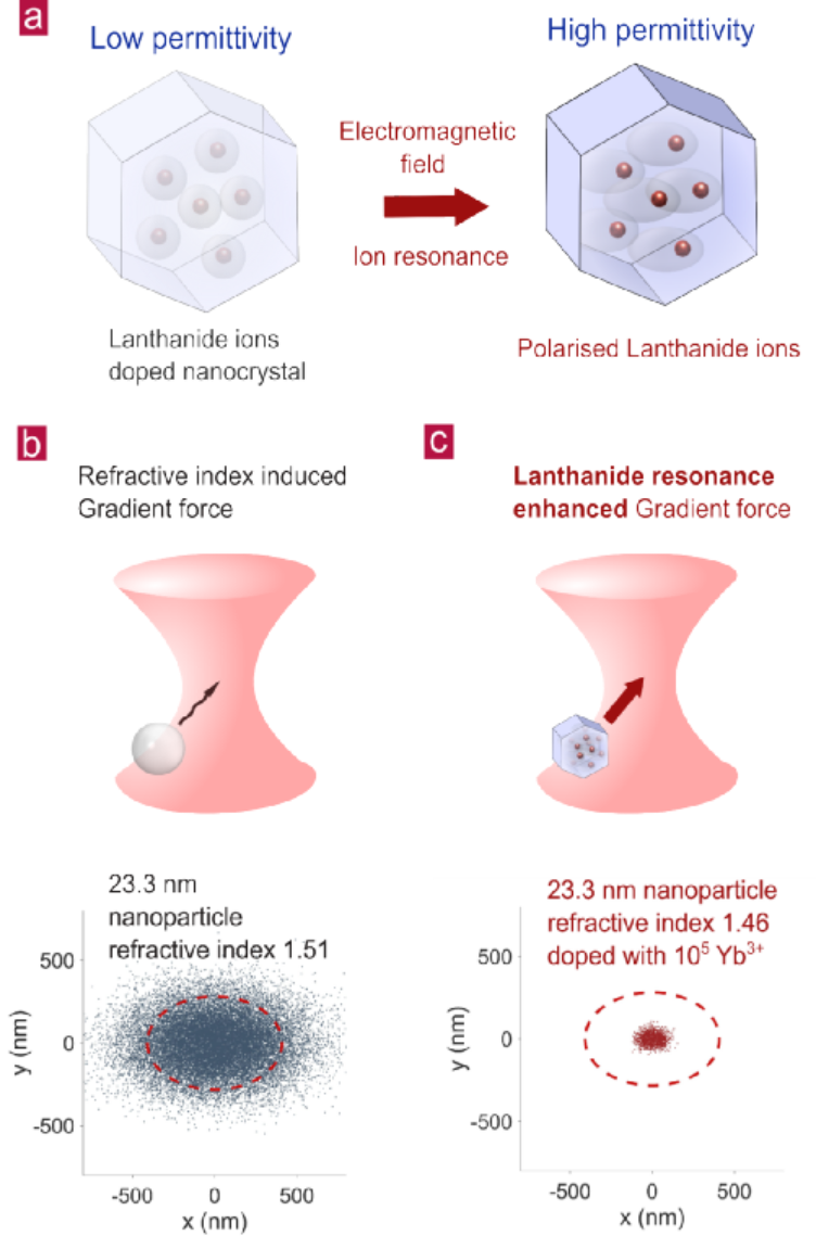
Fig. 1 | Comparison between optical trapping of low refractive index nanoparticles with or without doping by lanthanide ions. a).Diagram of the resonance effect for highly doped nanocrystals that up-regulates the permittivity and polarisability of a low index nanocrystal. Comparison of simulated 2D position distributions of a single nanoparticle optically trapped with b). conventional gradient force generated by refractive index mismatch and c). enhanced gradient force by lanthanide ions’ resonance effect. The power and wavelength of the trapping laser are 50 mW and 976.5 nm. The red circle indicates the spot size (1/e2 of intensity point) of an optical beam, 0.41 μm along the x-axis and 0.28 μm along the y-axis, respectively. The 2D position distributions are generated by a Monte Carlo simulation. The trap stiffnesses used to generate the distributions are simulated. The 2D distribution of experimental data with the same condition.
研究團隊首先對比了在相同的尺寸與折射率下,摻雜與未摻雜鑭系元素顆粒在光鑷捕捉情況下的二維位置分布模擬。從模擬結果可以發現,鑭系摻雜顆粒位置分布相比于未摻雜顆粒分布更密集,這是由于摻雜了鑭系元素的納米顆粒在976.5nm的捕獲激光下有更高的極化率,導致梯度力增強使得光鑷勢阱剛度更高。
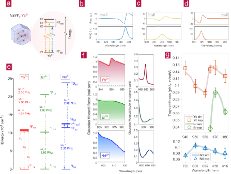
Fig. 2 | Investigation of ytterbium, erbium and neodymium doping in enhancing the optical gradient force. a). Illustration of energy levels of Yb3+ doped in NaYF4 nanocrystals. The calculated real and imaginary parts of electromagnetic susceptibility () for transitions b)E5-E1(
), c) E6-E1(
), and d) E7-E1(
). The concentration of Yb3+ resonator ions is 1.5 nm-3. e). Simplified energy level diagrams of Yb3+, Er3+ and Nd3+ in NaYF4 nanocrystal.
,
, and
are the dipole resonance angular frequencies. f). The calculated Clausius Mossotti factors (CM) for Yb3+, Er3+ and Nd3+ in NaYF4 crystal host. The real part of CM for pure NaYF4 crystals is shown as grey dash lines at a constant value of 0.064. g). The experimentally measured trap stiffness for Yb3+, Er3+ and Nd3+doped nanocrystals at different laser wavelengths. The longest trapping wavelength is 980nm due to the limited tuning range of trapping laser. The trapping range for Nd3+ is between 795 nm and 815 nm to generate sufficient emission intensity for trap stiffness measurement. The thicker shadowing lines are the theoretical simulation results based on the physical parameters of different nanoparticles.
通過模擬可以看出鐿離子(Yb3+)對于不同波長有不同的吸收曲線,鉺離子(Er3+)和釹離子(Nd3+)由于能級的不同也在對應波長有不同的吸收。這一特性導致了不同波長的捕獲激光會激發不同材料的極化率也不一樣,導致了同一中摻雜顆粒的捕獲力在波長變化時會有不同的捕獲力。
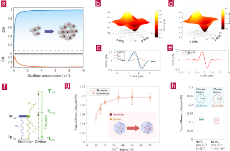
Fig. 3 | Effect of oscillated ions’ concentration on optical trapping. a). rCM and iCM for different oscillator concentration. The vertical dashed line shows the oscillators’ concentration ( = 1.5 nm-3) for the trapping power 50 mW. The horizontal dashed line shows the rCM for pure NaYF4 crystal. b). Axial force (Fz) distribution andd). lateral force (Fy) distribution in the y-z plane. The position of z = 0 indicates the force zero position.c). The y = 0 cross-line at Fz and e). the z = 0 cross-line at Fy.f). Diagram of distribution and transition of the excited state Yb3+ carriers. g).The experimentally measured trap stiffness for Yb3+doped nanoparticles, varying with doping concentration. h). The trap stiffness for different emitter (Er3+ and Tm3+) with the same sensitiser and emitter concentration. The median values of the box plot are 0.115 and 0.111 pN/μm/mW for Er3+ and Tm3+ doped nanoparticle, respectively.
通過課題組的模擬計算,可以通過鑭系元素的摻雜濃度優化上轉換納米顆粒的捕獲力,從而在相同尺寸的前提下提高捕獲力。在對應的諧振波長下,相同的鐿離子(Yb3+)摻雜濃度在相同尺寸下的捕獲力基本相同,與熒光離子的濃度無關。
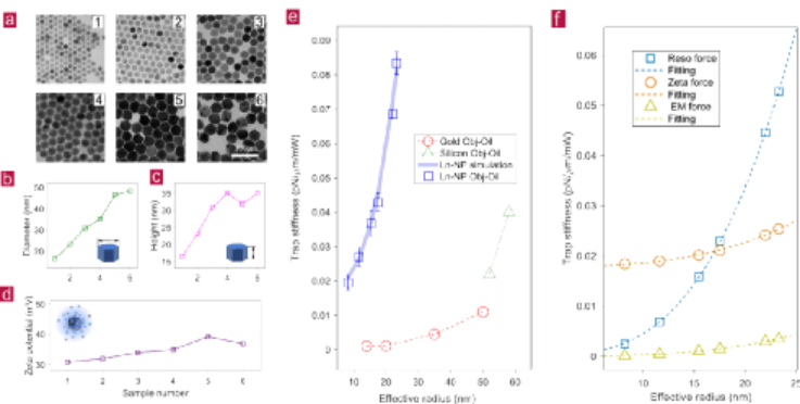
Fig. 4 | Trap stiffness measurements of lanthanide-doped nanoparticles at a different volume. a). TEM images ofsix typical batches ofNaYF4: 20% Yb3+, 2% Er3+ nanoparticles with different volume. The sizes of the particles are gradually increased from No.1 to No. 6, as shown in their b) averaged values of diagonal diameters and c) heights. d). The averaged values of zeta potentials of the above nanoparticles. e). The trap stiffness values of single lanthanide-doped nanoparticles. The effective radius is calculated from . For comparison, the lateral trap stiffness values of Gold and Silicon nanoparticles are quoted from previous reports by Oddershede’s group and Reece’s group, respectively. f). The calculated proportions of three factors contributing to the measured optical trapping force reported in e), ion-doping resonance-enhanced force (Reso force), zeta potential enhanced force (zeta force), and the classical electromagnetic force (EM force). All the experimental data has been collected by an oil immersion objective lens (Obj-Oil).
課題組還測試了相同鑭系摻雜濃度下(NaYF4: 20% Yb3+, 2% Er3+)不同體積上轉換納米顆粒在相同激光下的捕獲力。結果與模擬結果完全一直。課題組進而分析了在不同尺寸下不同種類的光力強度的比例,包括離子共振光力、表面電勢光力和傳統光力。當納米顆粒大于17納米時,這種新型的離子共振光力開始起主導效果。同時結果還展示出了25納米的低折射率的上轉換納米顆粒捕獲力比同體積的高折射率金納米顆粒還要高30倍。
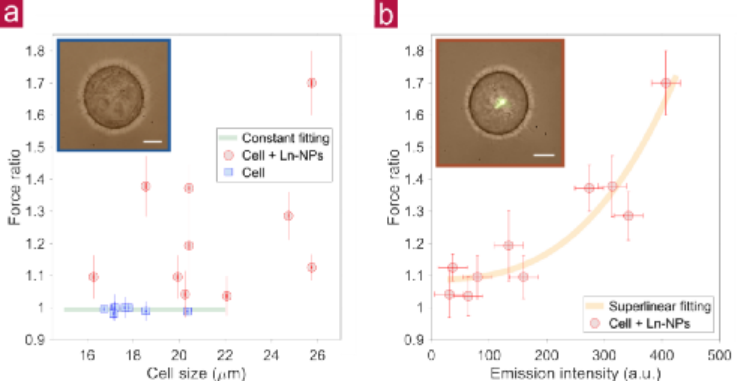
Fig. 5 | The escape velocity measurement to quantify the trap stiffness for Hela cells with and without lanthanide-doped nanoparticles (Ln-NPs). The ratio of the drag force by 976.5 nm laser to that by 808 nm laser (as control) shows the enhancement on the trap stiffness by lanthanide doped nanoparticles under the 976.5 nm trapping laser. The force ratio is obtained by measuring the ratio of escape velocity by 976.5 nm laser to that by 808 nm lase, as the force ratio value is equal to the ratio of escape velocity. a). The force ratios for seven randomly trapped Hela cells without nanoparticles (blue square dots) compared with the ten randomly trapped cells with nanoparticles (red circular dots). The insert figure shows one of the trapped cells without nanoparticle. b). The re-plot of force ratio of the ten nanoparticle-stained cells as a function of the upconversion emission intensity indicating the amount of nanoparticles. The insert figure shows one typical cell, by merging the bright field image and the fluorescence image excited by the 976.5 nm trapping laser. The Scale bar is 5 μm. The power of trapping laser at 976.5 nm and 800 nm are 90 mW and 88 mW, respectively. Each data point is averaged from four independent measurements.
最終研究團隊將上轉換納米顆粒綁定到海拉細胞表面,并且用光鑷抓取細胞在水中快速拖動觀測細胞的脫離速度是否因為綁定了上轉換顆粒而改變。結果表明,表面綁定了上轉換顆粒的細胞脫離速度更高,捕獲力更強,這一實驗為上轉換納米顆粒在生物力學應用方向上提供了新思路。
結論與展望
與其他納米顆粒相比,高摻雜的鑭系粒子在電磁場中有很強的諧振特性導致光鑷捕獲力顯著提升。這個技術使得納米顆粒在較小的折射率差溶液中的抓取成為了可能,同時更高的捕獲力可以提供更高精度的力學測量與應用。低折射率導致的低熱量吸收也打開了高效、長期利用光鑷抓取生物樣品的大門。未來,結合上轉換納米顆粒獨特的光學傳感特性與光鑷技術將可能實現細胞內部納米級別的溫度、pH和力的同時測量。本課題是課題組研究方向之一,其進展的順利主要得益于金大勇教授提供的高水平科研平臺與王帆博士的細心指導,該課題也得到了新南威爾士大學Peter Reece博士的鼎力支持。
參考文獻
Shan, X., Wang, F., Wang, D. et al. Optical tweezers beyond refractive index mismatch using highly doped upconversion nanoparticles. Nat. Nanotechnol. (2021).
DOI:10.1038/s41565-021-00852-0
https://doi.org/10.1038/s41565-021-00852-0
作者介紹

金大勇教授:金大勇,悉尼科技大學杰出教授和南方科技大學講席教授。2007年博士畢業于麥考瑞大學,2015年任悉尼科技大學教授,2017 年任杰出教授,2019年任南方科技大學講席教授。作為所長,他五年內先后組建了澳洲科研基金委資助的可集成生物醫學器件與技術轉化中心,中澳科學與研究基金資助的便攜式體外診斷技術聯合研究中心,悉尼科大-南科大生物醫學材料和器件聯合研究中心, 和悉尼科大生物醫學材料及儀器研究所。金大勇教授已發表SCI高水平論文150余篇、其中包括Nature及其子刊25篇。他的專業領域涵蓋了生物工程光學、納米探針技術、生物醫療診斷、精密光學儀器、微流控芯片等領域。并于2015年榮獲澳洲科研最高獎尤里卡獎交叉學科創新獎,2016年當選澳大利亞百名科技創新領軍人物,2017年榮獲澳洲科學院工程科學獎,以及2017年榮獲澳大利亞總理獎 - 年度理學家獎。https://www.wxwenku.com/d/108750009
生物醫學材料及儀器研究所主頁:http://ibmd.uts.edu.au/

王帆博士:王帆博士于2014年在澳洲新南威爾士大學獲得博士學位。博士期間,他致力于研究光鑷操作納米顆粒的研究。之后,王博士于2013年加入澳洲國立大學Prof Chennupati Jagadish (澳洲物理工程學院院士) 課題組,負責領導和管理光學方向。期間從事納米激光,二維材料以及凝聚態物理的研究。從2015年開始,王博士開始轉向生物光子學,加入了麥考瑞大學澳洲納米光子學國家研究中心,以及金大勇課題組。期間負責領導課題組的生物光子學方向,博士生導師。同年加入悉尼科技大學金大勇教授團隊,以及生物材料器件中心(IBMD), 繼續擔任生物光子學方向的負責人,博士生導師。并且開始幫助金大勇教授組建在悉尼科技大學的初始團隊。王博士于2020年在悉尼科技大學電子工程系成立課題組進行光鑷、激光制冷以及器件化超分辨成像技術的研究。王帆博士一共發表了SCI 論文62篇,其中包括Nature及其子刊13篇。
金大勇課題組以及王帆博士課題組誠招有從事生物光子學意向的優秀碩士以及博士學生。


